Abstract
The cementless Oxford unicompartmental knee replacement has been demonstrated to have superior fixation on radiographs and a similar early complication rate compared with the cemented version. However, a small number of cases have come to our attention where, after an apparently successful procedure, the tibial component subsides into a valgus position with an increased posterior slope, before becoming well-fixed. We present the clinical and radiological findings of these six patients and describe their natural history and the likely causes. Two underwent revision in the early post-operative period, and in four the implant stabilised and became well-fixed radiologically with a good functional outcome.
This situation appears to be avoidable by minor modifications to the operative technique, and it appears that it can be treated conservatively in most patients.
Cite this article: Bone Joint J 2014;96-B:345–9.
The Oxford Unicompartmental Knee Replacement (OUKR, Biomet, Bridgend, United Kingdom) has been used for over 30 years and has excellent results in large series.1-3 However, national joint registries (NJRs) demonstrate a significantly higher rate of revision for unicompartmental knee replacement (UKR) compared with total knee replacement (TKR).The latest report from the NJR for England and Wales reports revision rates of 11.6% and 3.1% at nine years for UKR and TKR respectively; aseptic loosening is the most common indication for revision.4,5 In most cases, UKR is performed using a minimally-invasive surgical (MIS) approach, which is associated with a shorter inpatient stay and faster recovery.6-9 However, the use of cement with this technique can be technically challenging and some failures are related to an inadequate cement mantle, impingement due to excessive cement, loose fragments of cement or the misinterpretation of radiolucent lines associated with cemented fixation.10,11 This led to the introduction of a cementless version of the OUKR, which was designed to eliminate these problems.
Cementless OUKR is associated with fewer radiolucencies than the cemented version, with similar functional outcome at five years.12 In centres where a large number of UKRs are performed, it has been shown to have an early survivorship which is similar to that of the cemented OUKR with no increase in the rate of complications.13,14 However, a small number of cases have come to our attention in which the tibial component of the implant subsides into a valgus position in the early post-operative period. Here, we describe the natural history, clinical and radiological findings of this subset of patients and describe their management.
Patients and Methods
The patients in this report underwent primary surgery at one of three institutions and came to the attention of the senior author (DWM) either through referral (internal or tertiary) or correspondence. They all underwent cementless OUKR for anteromedial osteoarthritis and, unless otherwise stated, conformed to the indications as defined by Goodfellow et al:15 full-thickness cartilage loss in the medial compartment with preservation of full-thickness cartilage in the lateral compartment and functionally intact cruciate and collateral ligaments. Surgery was performed by both experienced and inexperienced surgeons, and in all cases, a MIS technique was used without eversion or dislocation of the patella
Patient 1
A 79-year-old man underwent a cementless OUKR 18 months after a successful cemented, contralateral OUKR. At operation, he was found to be anterior cruciate ligament (ACL)-deficient, but the decision was made to persevere with UKR in view of his age, as the other conditions were met, and because of the success of the contralateral OUKR, which was also ACL deficient. At six weeks, there was an effusion with 5º to 10º of flexion deformity. This is not unusual at this stage following UKR; post-operative radiographs demonstrated no adverse features (Fig. 1a and 1d). He was mobile and comfortable and was referred for physiotherapy. At four months, having flexed the knee fully, he complained of pain deep within the knee, and radiographs showed that the tibial component had subsided from a position of 4º varus relative to the mechanical axis of the tibia to 8º of valgus and appeared loose (Fig. 1b and 1e). Revision of the tibial component was arranged. At the time of pre-operative assessment, the pain had improved and given that the implant had subsided no further, surgery was cancelled. By the end of the third post-operative year, the implant had stabilised and the Oxford Knee Score (OKS)16,17 was 39/48. His latest radiograph, at five years post-operatively, shows excellent fixation with no radiolucency (Fig. 1c and 1f). He has developed degenerative spinal disease and his OKS has declined (bilaterally) to 29/48.
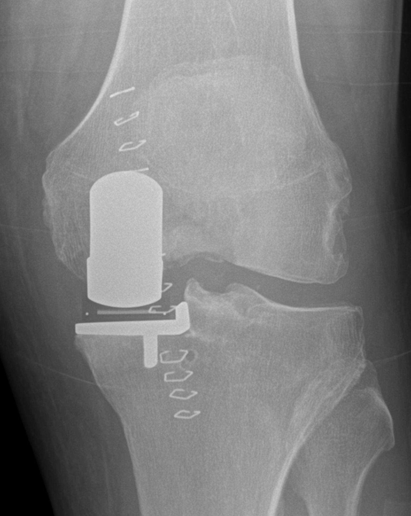
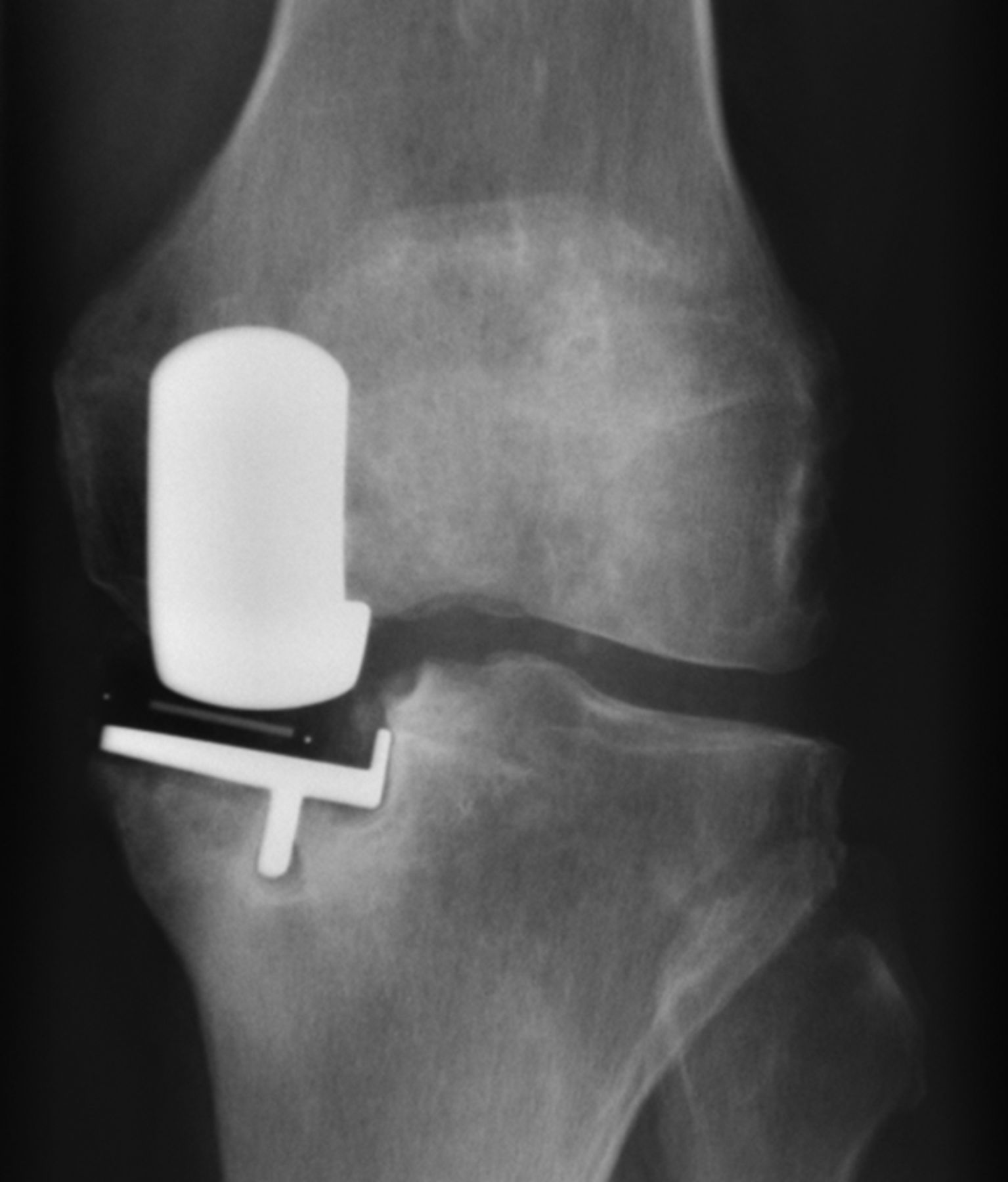
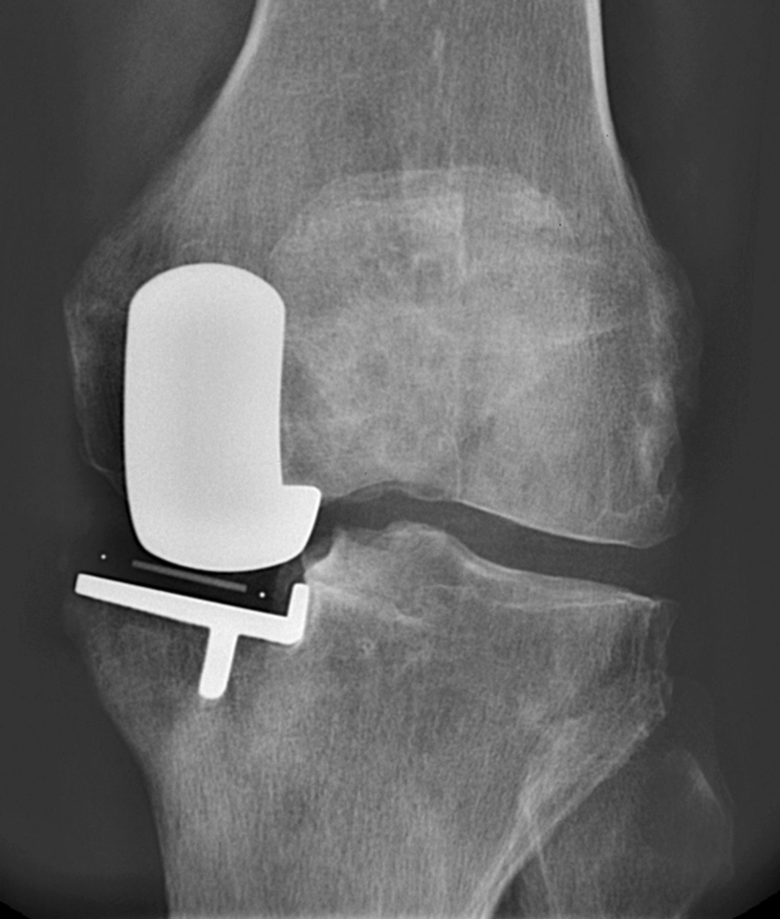
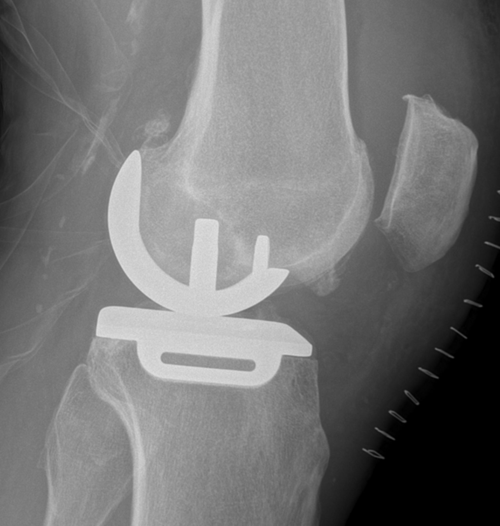
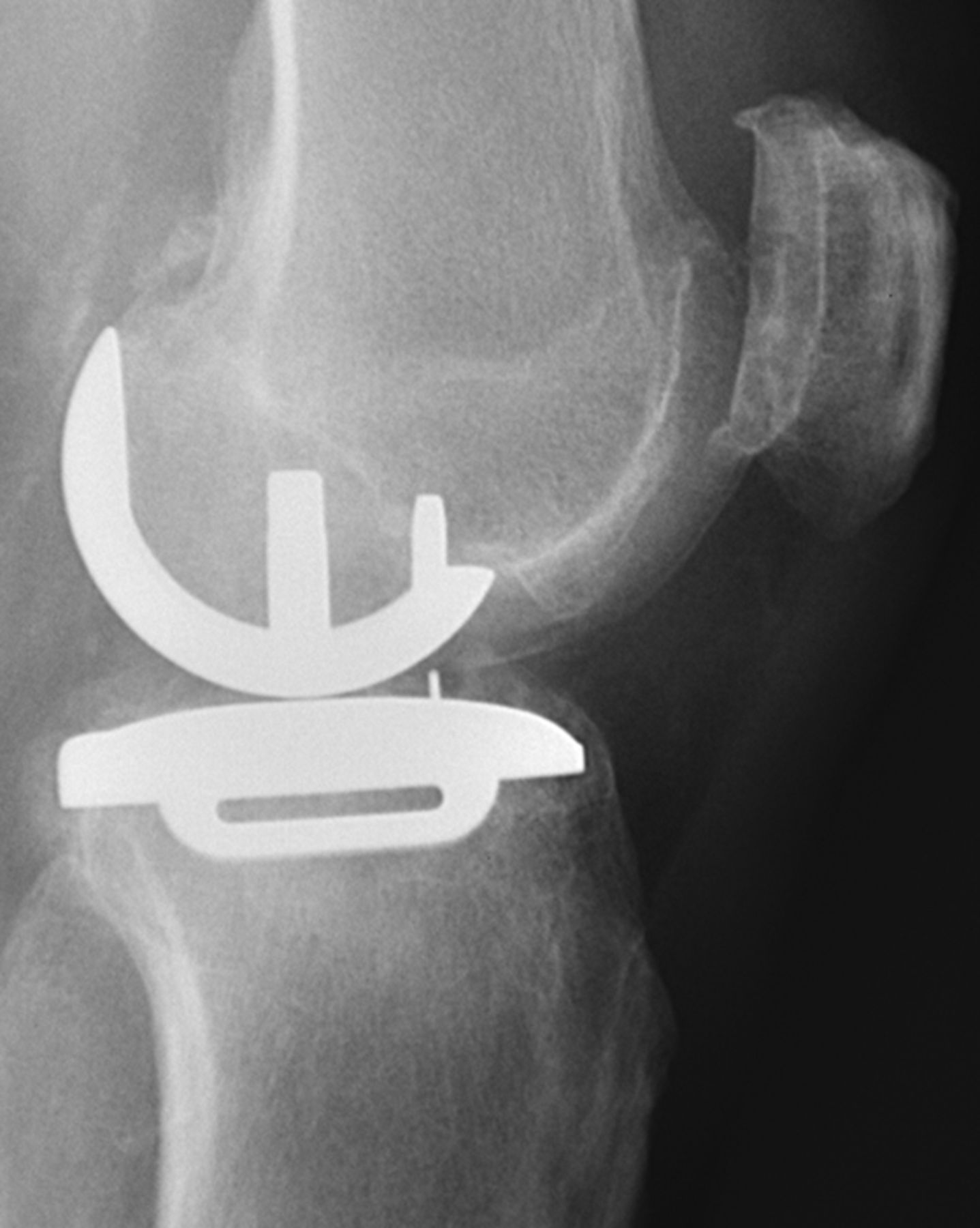
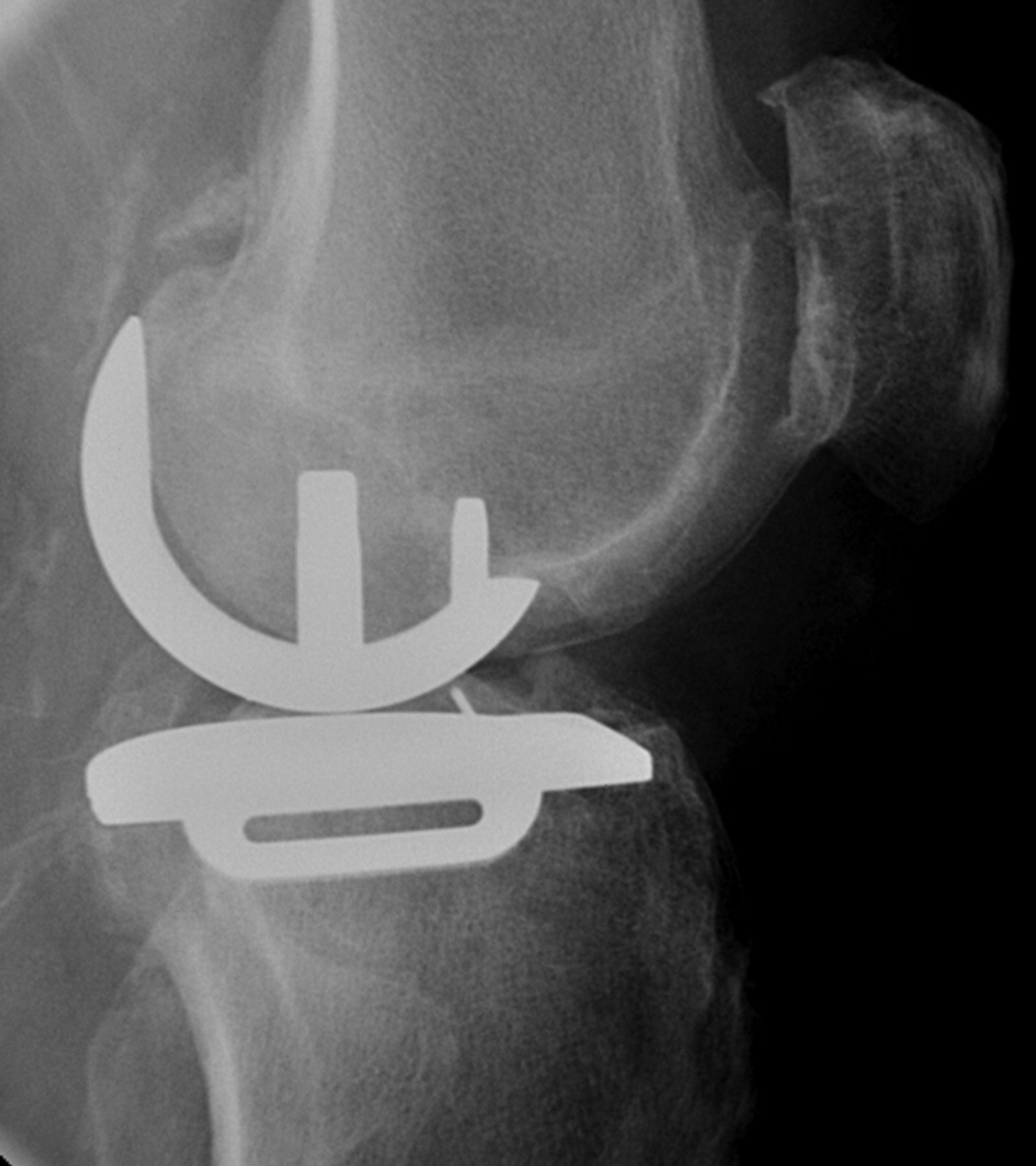
Figs. 1a - 1f
Radiographs showing a) anteroposterior view of patient 1 immediately post-operatively, b), following subsidence and c), five years post-operatively. Radiograph d) shows lateral view of patient 1 immediately post-operatively; e), following subsidence, and f) five years post-operatively.
Patient 2
A 66-year-old man underwent an uncomplicated cementless OUKR; there were no abnormal findings intra-operatively and the immediate post-operative radiograph showed that the implant was in a satisfactory position. In the fourth post-operative week, he developed increasing antero-medial pain, and radiographs showed subsidence of the tibial component into valgus, with lucency below the keel of the prosthesis. The patient was instructed to begin partial weight-bearing with crutches and was reviewed at eight weeks, at which time the pain had subsided and the component had stabilised. He has reported no further problems.
Patient 3
A 66-year-old man underwent an apparently successful OUKR with an uneventful first six post-operative weeks. Over the subsequent weeks, and without a clear precipitating factor, he developed further medial pain. A radiograph showed subsidence of the implant into valgus. The symptoms did not improve with partial weight-bearing and revision was undertaken, at which time the implant was found to be loose, with bone ingrowth restricted to the keel (Fig. 2). Revision to a cemented tibial component rendered him pain-free.
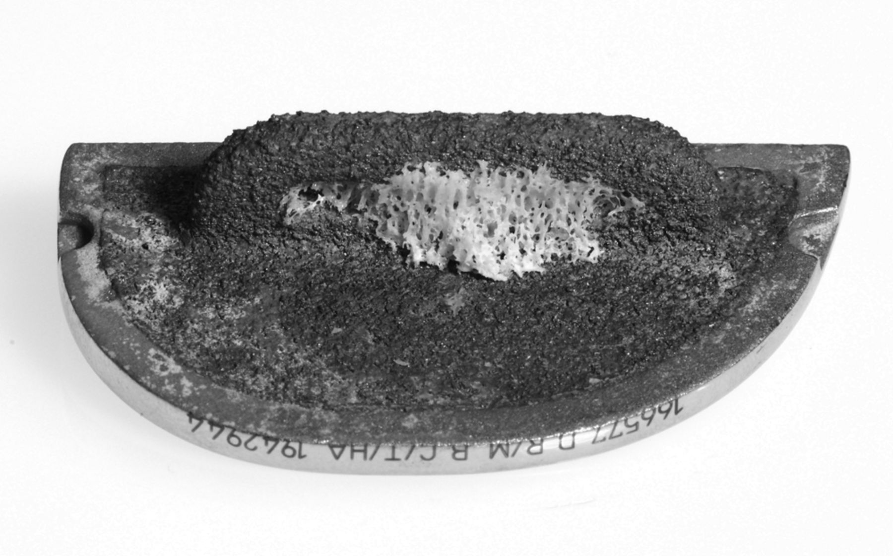
Fig. 2
Photograph of the explanted tibial component from patient 3, showing bony ingrowth around the keel.
Patient 4
A male patient underwent a cementless OUKR at the age of 65 years and had made a good immediate post-operative recovery but subsequently found the knee stiff, with difficulty achieving full extension. Over the following six months, the stiffness did not improve and the knee became progressively more painful. Review of immediate post-operative radiographs showed an under-sized tibial component in a good position. Subsequent radiographs demonstrated progressive subsidence into a valgus position, particularly of the posterior part of the component on the lateral radiograph (Fig. 3).
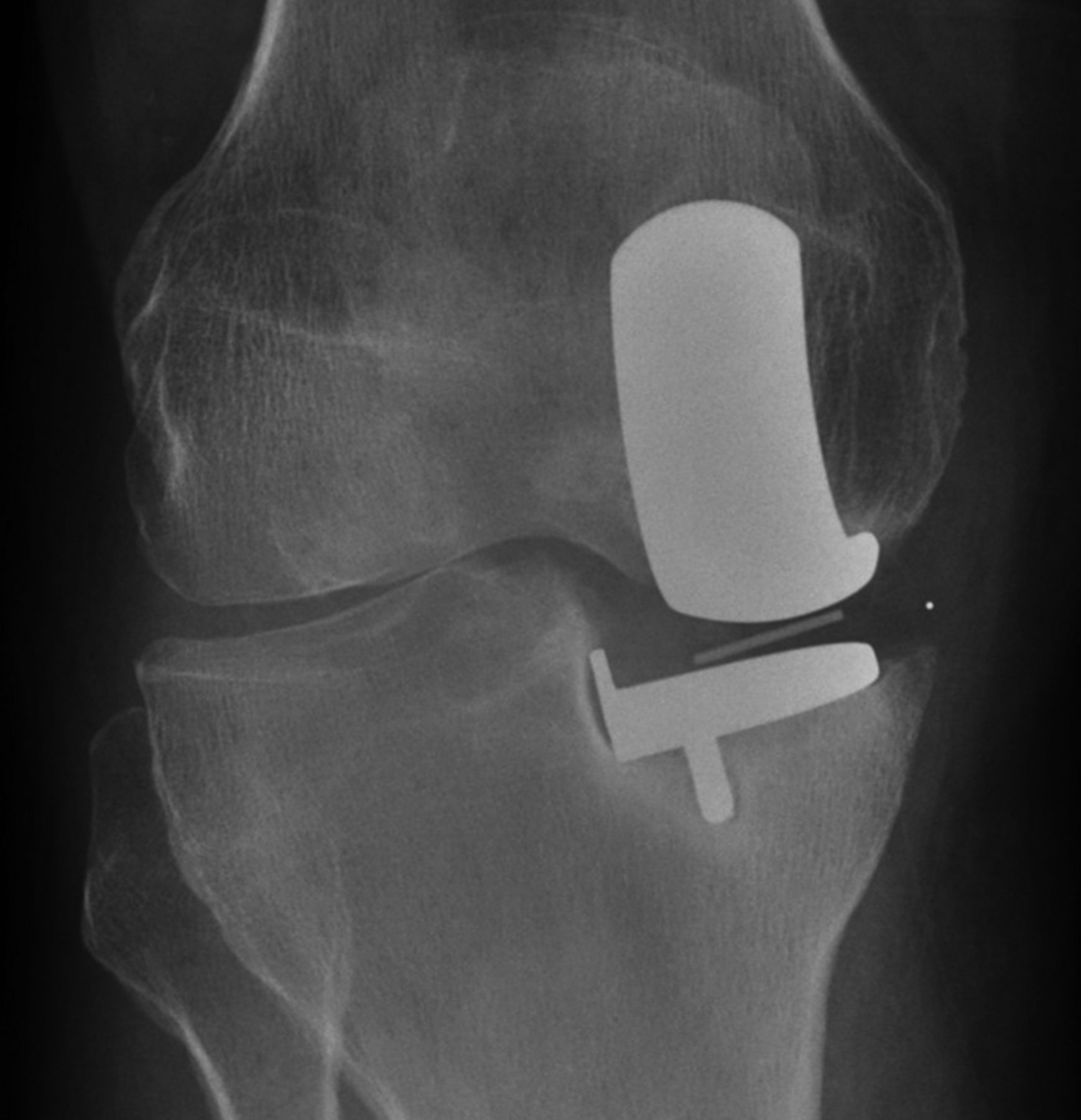
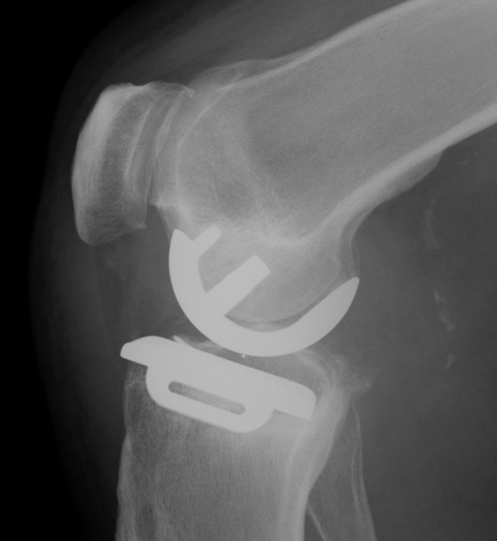
Figs. 3a - 3b
Radiographs showing a) anteroposterior and b) lateral views of patient 4, demonstrating valgus and posterior subsidence of an under-sized tibial component.
He underwent revision, at which time the tibial component was found to be well-fixed, with an un-displaced fracture of the posterior tibial cortex. He made an unremarkable recovery after revision to a TKR.
Patient 5
A 62-year-old woman underwent cementless OUKR and the first six post-operative weeks were uneventful. She presented again six months post-operatively, with increasing anteromedial tibial and femoral pain and reduced mobility. The radiograph demonstrated subsidence into valgus, with minimal increase in tibial slope. Exploration was undertaken 15 months post-operatively. Both components were found to be well-fixed but there was evidence of anterior femoral impingement. Debridement was undertaken with insertion of a 2 mm thicker bearing. At the latest follow-up, ten months after this second procedure, the symptoms have improved with an OKS of 35, which is a 21 point improvement on her pre-operative score.
Patient 6
A 76-year-old man underwent cementless OUKR, having previously undergone the same procedure on the other side successfully. Radiographs showed satisfactory alignment of the components. After two pain-free post-operative weeks, he developed severe anteromedial pain on weight-bearing. Radiographs taken three months post-operatively showed valgus–posterior subsidence. Revision was advised but by three months following primary surgery, there had been no further subsidence and there was improvement in symptoms. This continued, and at two years post-operatively, radiographs show a well-fixed component and the OKS was 36/48.
Discussion
While these six patients were treated at three separate institutions, their clinical and radiological findings are similar. All had a good early result from their OUKR before having further symptoms. All tibial components had tipped into a valgus position, with subsidence of the posterior part of the tibial tray, which appeared loose on radiographs.
In four patients, there was subsequent clinical improvement and radiological evidence of good fixation within the first post-operative year. It is now at least two years following the initial procedure for these patients and all have good radiological evidence of fixation and satisfactory outcome scores.
The remaining two were revised in the early post-operative period; one was found to be well-fixed and the other to have partial bony ingrowth around the keel. We do not know what would have happened had revision not been undertaken; the experience from the other patients presented here suggests that the symptoms are likely to have settled.
When determining the optimal management for similar patients, the risks of watchful waiting, which include the potential for bone loss that may complicate revision surgery, should be weighed against the risk of performing revision surgery which might be unnecessary. None of these patients had evidence of progressive bone loss; in fact, all show radiological evidence of secondary osseointegration with clinical improvement. Therefore we advise against early revision under these circumstances and suggest conservative management, with a period of partial weight-bearing and close clinical and radiological follow-up.
The explanation for this phenomenon is unclear. Although there was concern at the time that these UKRs might have been infected, none had symptoms or haematological evidence of infection. There was no radiological evidence of osteopenia in any of the patients. While two of the patients, aged 79 and 76 years respectively, are older than most in the NJR, in which the median age is 64 years (interquartile range 58 to 71),5 the demographic profile of these patients is similar to those in previous studies of cemented and cementless OUKR from specialist centres.3,14
Consideration of the pattern of subsidence and the natural history of this group of patients leads us to speculate that the appearances may result from impingement of the mobile bearing against the lateral wall of the tibial tray during flexion in the early post-operative period. The bearing tends to move away from the lateral wall in extension and therefore such impingement would not be detectable on an anteroposterior, standing radiograph. In normal circumstances without impingement, the centre of load lies within the central third of the tibial component in both the sagittal and coronal planes, and the load in the tibia beneath the component is entirely compressive. If the femoral component is positioned slightly too far laterally relative to the tibial component, impingement of the mobile bearing on the lateral wall may occur. This may result in subluxation of the femur in the bearing and subsequent tilting of the bearing relative to the tibial tray, which dramatically alters the loading of the tibial component. Excessive compressive loads will be present lateral to the tibial keel, with unloading of the medial part of the tibial component. These excessive loads, if applied prior to full integration of the tibial plateau, will cause the tibial component to subside into valgus. Once in this position, the bearing will no longer impinge on the wall and normal loading is restored (Fig. 4). The porous titanium and hydroxyapatite coating of the cementless implant allows bone ingrowth and fixation in this new position.
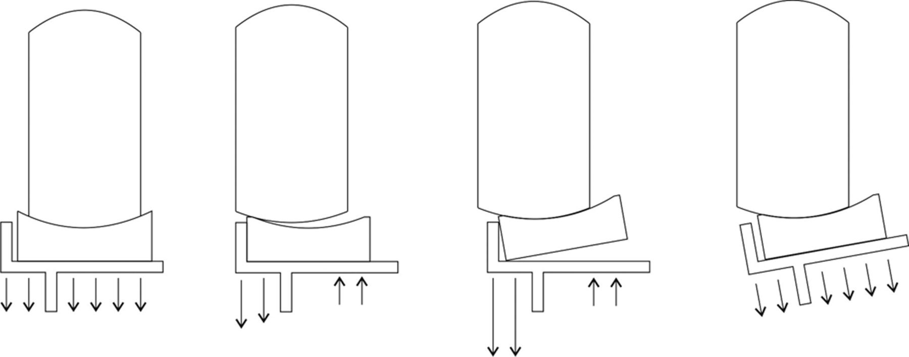
Fig. 4
Schematic demonstration of the possible mechanism. From left to right: Normally-functioning cementless OUKR. Relative lateralisation of the femur on the tibia causes the bearing to abut the lateral tibial wall. The femoral component subluxes on the bearing, and the bearing may tip in response. In this situation, the forces lateral to the keel become very large and there may be tension at the interface medially. As a result, the tibia subsides into valgus, in which case, the bearing will move away from the wall, normalising the loads beneath the tray and allowing secondary fixation.
Another factor that may predispose to collapse of the tibial component into valgus is damage to the lateral part of the horizontally-cut surface of the medial tibial plateau. This may occur if the vertical saw cut is too deep. Extensive damage may occur if the cut is initially too medial and another deep cut is performed more laterally, thus creating an area of unsupported bone. This can be prevented by performing the horizontal cut first (slightly undermining the tibial eminence) and inserting an ‘angel wing’ into the lateral side of the cut. The vertical cut can then be performed, with the ‘angel wing’ preventing it from going too deep.
All of these cases demonstrate some subsidence of the posterior part of the implant. In one of the revised cases (patient 4), there was evidence of a fracture of the posterior cortex at the time of revision, which is likely to have occurred at the time of primary surgery. This case appeared to be slightly under-sized and, as such, may be different from others. However, subsidence into an increased posterior slope was a universal finding and some of these cases may represent occult posterior cortical fractures.
Factors predisposing to periprosthetic fracture include deepening the posterior cortical sagittal saw cut during implantation,18 damage to the posterior cortex during curettage of the keel cut,19 failure to fully clear the keel cut and excessive force of impaction. The cementless OUKR may be more sensitive to these errors than the cemented version.18 In order to avoid these errors, we recommend that the specific keel-cutting saw is always used and only through the template, rather than directly onto the cut tibial surface. The template-fixing pin should be held in place by the surgeon. A trial tibial component should be inserted and the keel-clearing pick should only be used if the trial component will not seat. If the pick is used, the front and back of the keel slot should be cleared through the template. Finally, only a light hammer should be used for impaction. If the component does not fully seat despite full clearance of the slot, this position should be accepted rather than persevering with impaction using excessive force.
The cementless OUKR has excellent short-term results.12-14 A study using radiostereometric analysis of this prosthesis has demonstrated minimal tibial migration with stabilisation in the first year,20 and a second study using iDXA has demonstrated preservation of bone mineral density adjacent to the tibial component.21 Cases of the type described here are rare: in a report of the first 1000 cementless OUKRs implanted in three centres, three patients had similar subsidence, although none to the same extent as those reported here, and none had significant symptoms.14 They also appear to follow a benign course with conservative management. Further research is necessary to assess the relative contribution of technique, implant design and the indications for operation to the development of this situation.
1 Lisowski LA , van den BekeromMP, PilotP, van DijkCN, LisowskiAE. Oxford Phase 3 unicompartmental knee arthroplasty: medium-term results of a minimally invasive surgical procedure. Knee Surg Sports Traumatol Arthrosc2011;19:277–284.CrossrefPubMed Google Scholar
2 Price AJ , SvardU. A second decade lifetable survival analysis of the Oxford unicompartmental knee arthroplasty. Clin Orthop Relat Res2011;469:174–179.CrossrefPubMed Google Scholar
3 Pandit H , JenkinsC, GillHS, et al.Minimally invasive Oxford phase 3 unicompartmental knee replacement: results of 1000 cases. J Bone Joint Surg [Br]2011;93-B:198–204.CrossrefPubMed Google Scholar
4 No authors listed. Australian Orthopaedic Association National Joint Replacement Registry Annual Report, 2013. https://aoanjrr.dmac.adelaide.edu.au/ (date last accessed 6 December 2013). Google Scholar
5 No authors listed. National Joint Registry for England, Wales and Northern Ireland: 10th Annual Report, 2013. http://www.njrcentre.org.uk/ (date last accessed 6 December 2013). Google Scholar
6 Lombardi AV Jr , BerendKR, WalterCA, Aziz-JacoboJ, CheneyNA. Is recovery faster for mobile-bearing unicompartmental than total knee arthroplasty?Clin Orthop Relat Res2009;467:1450–1457.CrossrefPubMed Google Scholar
7 Hopper GP , LeachWJ. Participation in sporting activities following knee replacement: total versus unicompartmental. Knee Surg Sports Traumatol Arthrosc2008;16:973–979.CrossrefPubMed Google Scholar
8 Brown NM , ShethNP, DavisK, et al.Total knee arthroplasty has higher postoperative morbidity than unicompartmental knee arthroplasty: a multicenter analysis. J Arthroplasty2012;27-S8:86–90.CrossrefPubMed Google Scholar
9 Kasch R , MerkS, DrescherW, et al.Marginal contribution of UKS- versus TKA in varus arthritis of the knee. Arch Orthop Trauma Surg2012;132:1165–1172.CrossrefPubMed Google Scholar
10 Dervin GF , CarruthersC, FeibelRJ, et al.Initial experience with the oxford unicompartmental knee arthroplasty. J Arthroplasty2011;26:192–197.CrossrefPubMed Google Scholar
11 Kendrick BJ , LonginoD, PanditH, et al.Polyethylene wear in Oxford unicompartmental knee replacement: a retrieval study of 47 bearings. J Bone Joint Surg [Br]2010;92-B:367–373.CrossrefPubMed Google Scholar
12 Pandit H , LiddleAD, KendrickBJ, et al.Improved fixation in cementless unicompartmental knee replacement: five-year results of a randomized controlled trial. J Bone Joint Surg [Am]2013;95-A:1365–1372.CrossrefPubMed Google Scholar
13 Hooper GJ , MaxwellAR, WilkinsonB, et al.The early radiological results of the uncemented Oxford medial compartment knee replacement. J Bone Joint Surg [Br]2012;94-B:334–338.CrossrefPubMed Google Scholar
14 Liddle AD , PanditH, O'BrienS, et al.Cementless fixation in Oxford unicompartmental knee replacement: a multicentre study of 1000 knees. Bone Joint J2013;95-B:181–187.CrossrefPubMed Google Scholar
15 Goodfellow J, O'Connor J, Dodd C, Murray D. Unicompartmental arthroplasty with the Oxford knee. Oxford: Oxford University Press, 2006. Google Scholar
16 Dawson J , FitzpatrickR, MurrayD, CarrA. Questionnaire on the perceptions of patients about total knee replacement. J Bone Joint Surg [Br]1998;80-B:63–69.CrossrefPubMed Google Scholar
17 Murray DW , FitzpatrickR, RogersK, et al.The use of the Oxford hip and knee scores. J Bone Joint Surg [Br]2007;89-B:1010–1014. Google Scholar
18 Seeger JB , HaasD, JagerS, et al.Extended sagittal saw cut significantly reduces fracture load in cementless unicompartmental knee arthroplasty compared to cemented tibia plateaus: an experimental cadaver study. Knee Surg Sports Traumatol Arthrosc2012;26:1087–1091.CrossrefPubMed Google Scholar
19 Sloper PJ , HingCB, DonellST, GlasgowMM. Intra-operative tibial plateau fracture during unicompartmental knee replacement: a case report. Knee2003;10:367–369.CrossrefPubMed Google Scholar
20 Kendrick BJ , BottomleyNJ, GillHS, et al.A randomised controlled trial of cemented versus cementless fixation in Oxford unicompartmental knee replacement in the treatment of medial gonoarthrosis using radiostereometric analysis. Osteoarthritis and Cartilage2012;20-S1:S36–S37. Google Scholar
21 Hooper GJ , GilchristN, MaxwellR, et al.The effect of the Oxford uncemented medial compartment arthroplasty on the bone mineral density and content of the proximal tibia. Bone Joint J2013;95-B:1480–1483.CrossrefPubMed Google Scholar
A.D. Liddle has been supported by research fellowships from Arthritis Research UK (grant number 20499) and the Royal College of Surgeons of England. D. W. Murray is supported by an NIHR Senior Research Fellowship.
The author or one or more of the authors have received or will receive benefits for personal or professional use from a commercial party related directly or indirectly to the subject of this article. In addition, benefits have been or will be directed to a research fund, foundation, educational institution, or other non-profit organisation with which one or more of the authors are associated.
This article was primary edited by D. Rowley and first proof edited by J. Scott.


